Ch. 3 - Observing Microorganisms Through a Microscope
Tortora - Microbiology: An Introduction 14th Edition
Tortora14th EditionMicrobiology: An IntroductionISBN: 9780138200398Not the one you use?Change textbook
Chapter 3, Problem 8

Assume that you are viewing a Gram-stained field of red cocci and blue rods through the microscope. You can safely conclude that you have
a. Made a mistake in staining
b. Two different species
c. Old bacterial cells
d. Young bacterial cells
e. None of the above

Verified step by step guidance
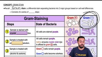
1
Step 1: Understand the Gram stain principle. Gram-positive bacteria retain the crystal violet stain and appear purple or blue, while Gram-negative bacteria do not retain the crystal violet and instead take up the counterstain (usually safranin), appearing red or pink.
Step 2: Identify the morphology and color described. Red cocci are spherical bacteria that appear red, indicating they are Gram-negative cocci. Blue rods are rod-shaped bacteria that appear blue, indicating they are Gram-positive rods.
Step 3: Recognize that different shapes and different Gram stain colors suggest the presence of more than one bacterial type or species in the sample.
Step 4: Consider that a single species typically has a consistent Gram reaction and morphology, so seeing both red cocci and blue rods means you are likely observing two different species.
Step 5: Conclude that the correct interpretation is that you have two different species present, corresponding to option (b).

Verified video answer for a similar problem:

This video solution was recommended by our tutors as helpful for the problem above.
Video duration:
2m
Was this helpful?

Key Concepts

Here are the essential concepts you must grasp in order to answer the question correctly.

Gram Staining and Color Interpretation

Gram staining differentiates bacteria into Gram-positive (purple/blue) and Gram-negative (pink/red) based on cell wall structure. Gram-positive bacteria retain crystal violet dye, appearing blue or purple, while Gram-negative bacteria take up the counterstain safranin, appearing red or pink.
Recommended video:

Bacterial Morphology

Bacteria are classified by shape, such as cocci (spherical) and rods (bacilli). Observing different shapes in the same field suggests the presence of multiple bacterial types or species, as morphology is a key identifying feature.
Recommended video:
Guided course
02:44
Types of Bacterial Cell Morphology

Interpreting Mixed Staining Results

Seeing red cocci and blue rods together usually indicates two different bacterial species with distinct Gram reactions. This is not typically due to staining error or cell age, but rather a mixed culture or sample containing diverse bacteria.
Recommended video:
Guided course
02:10
Introduction to Staining