Textbook Question
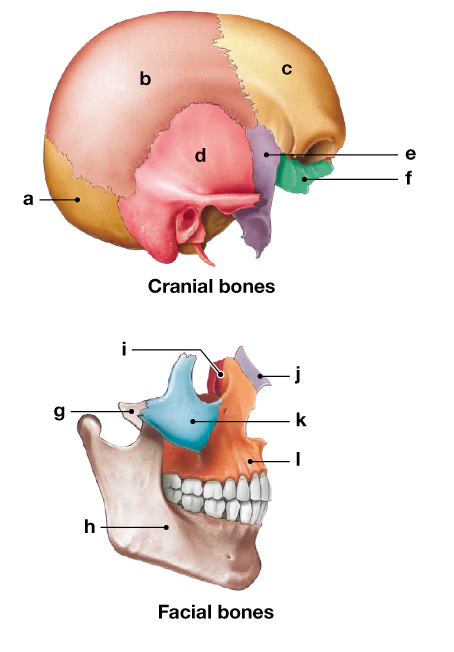
The unpaired facial bones include the:
(a) Lacrimal and nasal
(b) Vomer and mandible
(c) Maxilla and mandible
(d) Zygomatic and palatine
1
views

 Verified step by step guidance
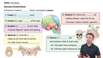
 8:20
8:20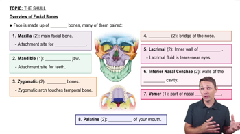 6:30
6:30 2:37
2:37The unpaired facial bones include the:
(a) Lacrimal and nasal
(b) Vomer and mandible
(c) Maxilla and mandible
(d) Zygomatic and palatine
Which of the following lists contains only facial bones? (a) mandible, maxilla, nasal, zygomatic, (b) frontal, occipital, zygomatic, parietal, (c) occipital, sphenoid, temporal, lacrimal, (d) frontal, parietal, occipital, sphenoid.
The boundaries between skull bones are immovable joints called:
(a) Foramina
(b) Fontanelles
(c) Lacunae
(d) Sutures