Skip to main content
Microbiology
My Course
Learn
Exam Prep
AI Tutor
Study Guides
Textbook Solutions
Flashcards
Explore
Try the app
My Course
Learn
Exam Prep
AI Tutor
Study Guides
Textbook Solutions
Flashcards
Explore
Try the app
Back
Reviewing the Different Types of Microscopes definitions
You can tap to flip the card.
Bright Field Microscope
You can tap to flip the card.
👆
Bright Field Microscope
Most common light microscope; observes stained or unstained specimens on a bright background, but may have poor contrast for transparent cells.
Track progress
Control buttons has been changed to "navigation" mode.
1/14
Related flashcards
Related practice
Recommended videos
Reviewing the Different Types of Microscopes quiz
Reviewing the Different Types of Microscopes
15 Terms
Reviewing the Different Types of Microscopes
9. Microscopes
4 problems
Topic
Nicole
Introduction to Staining
9. Microscopes
Topic
9. Microscopes - Part 1 of 3
6 topics
12 problems
Chapter
Nicole
9. Microscopes - Part 2 of 3
8 topics
10 problems
Chapter
Nicole
9. Microscopes - Part 3 of 3
1 topic
3 problems
Chapter
Guided course
05:32
Reviewing the Different Types of Microscopes
3
views
Terms in this set (14)
Hide definitions
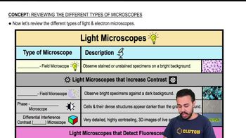
Bright Field Microscope
Most common light microscope; observes stained or unstained specimens on a bright background, but may have poor contrast for transparent cells.
Compound Light Microscope
A type of bright field microscope with multiple lenses, allowing calculation of total magnification for specimen observation.
Dark Field Microscope
Produces a dark background, making specimens appear brighter and enhancing contrast for better visualization.
Phase Contrast Microscope
Enhances visibility of dense cell structures, making them appear darker against a grayish background without staining.
Differential Interference Contrast Microscope
Generates highly detailed, three-dimensional images of live specimens with enhanced contrast.
Confocal Scanning Laser Microscope
Provides high contrast and three-dimensional imaging by focusing on multiple planes within a specimen.
Two-Photon Microscope
Enables high contrast, three-dimensional, and time-lapse imaging of deep structures, similar to x-ray vision.
Super Resolution Microscope
Offers extremely high resolution, around 0.01 micrometers, surpassing standard light microscopes.
Electron Microscope
Uses electrons instead of visible light to magnify specimens, allowing for much higher resolution imaging.
Transmission Electron Microscope
Creates two-dimensional images by passing electrons through specimens, ideal for visualizing internal cell structures.
Scanning Electron Microscope
Generates three-dimensional images by detecting electrons scattered from specimen surfaces, revealing external structures.
Resolution
The ability to distinguish fine details and structures, with higher values allowing clearer, more detailed images.
Contrast
The difference in visual properties that makes an object distinguishable from its background, crucial for specimen visualization.
Magnification
The process of enlarging the appearance of a specimen, allowing for detailed observation of microscopic structures.
 BackBack
 05:32
05:32