 Back
BackA Tour of the Cell: Prokaryotic and Eukaryotic Cell Structure and Function
Study Guide - Smart Notes
 Tailored notes based on your materials, expanded with key definitions, examples, and context.
Cell Types and Their Organization
Prokaryotic vs. Eukaryotic Cells
Cells are the fundamental units of life, and they are classified into two main types: prokaryotic and eukaryotic. Understanding their differences is essential for studying cell biology.
Prokaryotic Cells: Small, simple cells lacking membrane-bound organelles. Their DNA is located in a region called the nucleoid.
Eukaryotic Cells: Larger, more complex cells with membrane-bound organelles, including a nucleus that houses their DNA. Found in animals, plants, fungi, and protists.
Prokaryotic Cell Structure
Main Features of Prokaryotic Cells
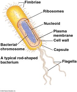
Prokaryotic cells, such as bacteria, have a simple structure but are highly efficient and diverse.
Nucleoid: Region where the cell's DNA is located (not enclosed by a membrane).
Ribosomes: Sites of protein synthesis.
Plasma Membrane: Selectively permeable barrier that encloses the cytoplasm.
Cell Wall: Rigid structure providing support and protection; composed of peptidoglycan in bacteria.
Capsule: Outer jelly-like layer for protection and adherence (in some bacteria).
Fimbriae: Hair-like structures for attachment to surfaces.
Flagella: Long, whip-like appendages for locomotion.
Eukaryotic Cell Structure
Overview of Eukaryotic Cells
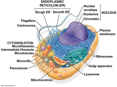
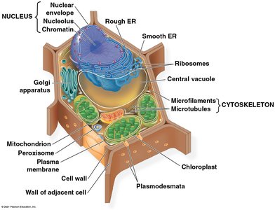
Eukaryotic cells are compartmentalized by internal membranes, allowing for specialized functions within organelles.
Nucleus: Contains the cell's genetic material (DNA) and is surrounded by a double membrane (nuclear envelope).
Nucleolus: Dense region within the nucleus; site of ribosomal RNA (rRNA) synthesis and ribosome assembly.
Chromatin: DNA-protein complex that organizes genetic material within the nucleus.
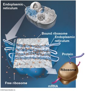
Ribosomes: Protein synthesis machinery, found free in the cytosol or bound to the endoplasmic reticulum (ER).
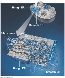
Endoplasmic Reticulum (ER): Network of membranes involved in protein and lipid synthesis.
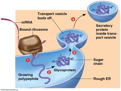
Rough ER: Studded with ribosomes; synthesizes proteins for secretion or membrane insertion.
Smooth ER: Lacks ribosomes; synthesizes lipids and detoxifies certain chemicals.
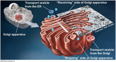
Golgi Apparatus: Modifies, sorts, and packages proteins and lipids for delivery to different destinations.
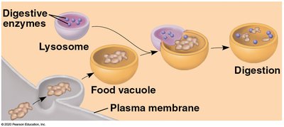
Lysosomes: Membranous sacs containing digestive enzymes for breaking down macromolecules and cellular debris.
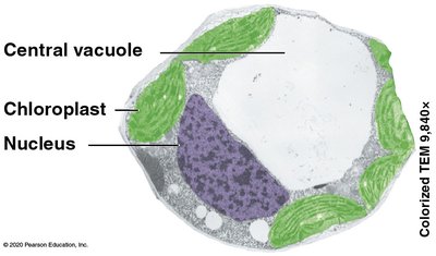
Vacuoles: Large vesicles for storage and transport; central vacuole in plants maintains turgor pressure.
Mitochondria: Sites of cellular respiration, generating ATP from nutrients.
Chloroplasts: Found in plants and algae; sites of photosynthesis.
Cytoskeleton: Network of protein filaments (microtubules, intermediate filaments, microfilaments) providing structural support and facilitating movement.
Plasma Membrane: Phospholipid bilayer controlling entry and exit of substances.
Plant vs. Animal Cells
While both plant and animal cells are eukaryotic, they have distinct features:
Plant Cells: Have a cell wall (cellulose), chloroplasts, and a large central vacuole.
Animal Cells: Lack cell walls and chloroplasts, but have centrioles and lysosomes.
The Nucleus and Ribosomes
Nucleus
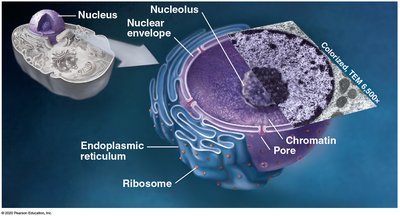
The nucleus is the control center of the cell, containing most of the genetic material.
Nuclear Envelope: Double membrane with pores for molecular exchange.
Nucleolus: Site of rRNA synthesis and ribosome assembly.
Chromatin: DNA wrapped around histone proteins.
Ribosomes
Ribosomes are the molecular machines that translate genetic information into proteins.
Free Ribosomes: Synthesize proteins for use within the cytosol.
Bound Ribosomes: Attached to the rough ER; synthesize proteins for secretion or membrane insertion.
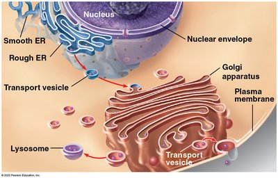
The Endomembrane System
Components and Functions
The endomembrane system is a group of interconnected organelles that work together to modify, package, and transport lipids and proteins.
Nuclear Envelope
Endoplasmic Reticulum (ER): Rough and smooth types
Golgi Apparatus
Lysosomes
Vesicles
Plasma Membrane
Endoplasmic Reticulum (ER)
The ER is a network of membranes involved in protein and lipid synthesis.
Rough ER: Studded with ribosomes; synthesizes proteins for export or membrane insertion.
Smooth ER: Synthesizes lipids, metabolizes carbohydrates, detoxifies drugs and poisons.
Protein Production in the ER
Proteins synthesized by ribosomes on the rough ER are processed and transported in vesicles to the Golgi apparatus for further modification and sorting.
Golgi Apparatus
The Golgi apparatus modifies, sorts, and packages proteins and lipids for secretion or delivery to other organelles.
Receiving Side (cis face): Accepts vesicles from the ER.
Shipping Side (trans face): Dispatches vesicles to their destinations.
Lysosomes
Lysosomes are membrane-bound organelles containing hydrolytic enzymes for intracellular digestion.
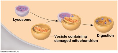
Digest macromolecules, old organelles, and foreign substances.
Play a key role in autophagy and defense against pathogens.
Vacuoles
Vacuoles are large vesicles with diverse functions, especially prominent in plant cells.
Central Vacuole: Maintains cell turgor, stores nutrients and waste products in plant cells.
Food Vacuoles: Formed by phagocytosis in some protists and animal cells.
Energy-Transforming Organelles
Mitochondria
Mitochondria are the powerhouses of the cell, generating ATP through cellular respiration.
Outer Membrane: Smooth and protective.
Inner Membrane: Folded into cristae to increase surface area for ATP production.
Matrix: Contains enzymes, mitochondrial DNA, and ribosomes.
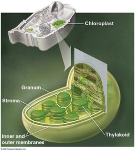
Chloroplasts
Chloroplasts are the sites of photosynthesis in plants and algae.
Thylakoids: Flattened sacs where light-dependent reactions occur.
Granum: Stack of thylakoids.
Stroma: Fluid surrounding thylakoids, contains DNA, ribosomes, and enzymes.
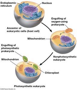
Endosymbiotic Theory
This theory proposes that mitochondria and chloroplasts originated as free-living prokaryotes that were engulfed by ancestral eukaryotic cells, forming a symbiotic relationship.
Evidence: Both organelles have their own DNA, double membranes, and ribosomes similar to prokaryotes.
Cytoskeleton and Cell Movement
Cytoskeleton
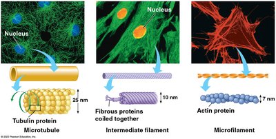
The cytoskeleton is a dynamic network of protein filaments that provides structural support, maintains cell shape, and enables movement.
Microtubules: Hollow tubes made of tubulin; involved in cell shape, organelle movement, and chromosome separation.
Intermediate Filaments: Rope-like fibers providing mechanical strength.
Microfilaments: Thin filaments of actin; involved in cell movement and muscle contraction.
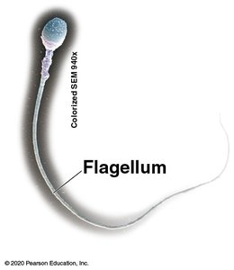
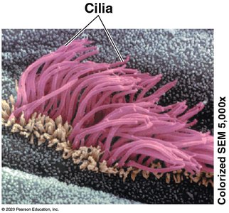
Cilia and Flagella
Cilia and flagella are motile appendages powered by the cytoskeleton, enabling movement or fluid transport across cell surfaces.
Cilia: Short, numerous, move substances past the cell.
Flagella: Longer, usually one or a few per cell, propel the cell itself.
Cell Surfaces and Junctions
Extracellular Matrix (ECM)
The ECM is a network of glycoproteins, polysaccharides, and proteoglycans outside animal cells, providing structural support and mediating cell signaling.
Collagen: Main structural protein in connective tissues.
Integrins: Transmembrane proteins connecting ECM to the cytoskeleton.
Cell Junctions
Cells are connected by specialized junctions that facilitate communication and maintain tissue integrity.
Junction Type | Structure | Function |
|---|---|---|
Tight Junction | Continuous seal between cells | Prevents leakage of extracellular fluid |
Desmosome | Anchoring junction using intermediate filaments | Fastens cells together |
Gap Junction | Cytoplasmic channels between cells | Allows communication and exchange of materials |
Plant Cell Walls and Plasmodesmata
Plant cells have a rigid cell wall made of cellulose, providing protection and structural support. Plasmodesmata are channels that connect the cytoplasm of adjacent plant cells, allowing for transport and communication.
Cell Wall: Composed mainly of cellulose, maintains shape, and prevents excessive water uptake.
Plasmodesmata: Openings in the cell wall for cytoplasmic exchange.
Summary Table: Prokaryotic vs. Eukaryotic Cells
Feature | Prokaryotic Cell | Eukaryotic Cell |
|---|---|---|
Nucleus | Absent | Present |
Membrane-bound Organelles | Absent | Present |
Cell Size | Small (1-10 µm) | Larger (10-100 µm) |
Examples | Bacteria, Archaea | Animals, Plants, Fungi, Protists |
