 Back
BackA Tour of the Cell: Prokaryotic and Eukaryotic Cell Structure and Function
Study Guide - Smart Notes
 Tailored notes based on your materials, expanded with key definitions, examples, and context.
Cell Components
Prokaryotic vs. Eukaryotic Cells
Cells are the fundamental units of life, and they are classified into two main types: prokaryotic and eukaryotic. Understanding their differences is essential for studying cell biology.
Prokaryotic Cells: Smaller, simpler, lack membrane-bound organelles, and do not have a true nucleus. DNA is located in a region called the nucleoid.
Eukaryotic Cells: Larger, more complex, contain membrane-bound organelles, and have a nucleus that houses their DNA.


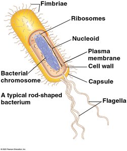
Prokaryotic Cell Structure
Prokaryotic cells, such as bacteria, have a simple structure but are highly efficient. Their main features include:
Nucleoid: Region where the cell's DNA is located (not enclosed by a membrane).
Ribosomes: Sites of protein synthesis.
Plasma Membrane: Selectively permeable barrier that encloses the cytoplasm.
Cell Wall: Provides structural support and protection.
Capsule: Jelly-like outer coating (in some bacteria) for protection and adherence.
Fimbriae: Hair-like structures for attachment to surfaces.
Flagella: Long, whip-like appendages for movement.
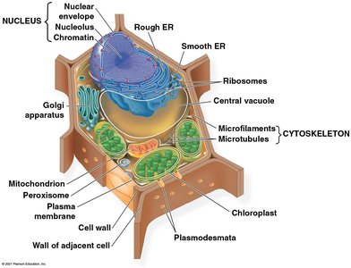
Eukaryotic Cell Structure
Eukaryotic cells are more complex and compartmentalized, allowing for specialized functions within organelles. They include animal, plant, fungal, and protist cells.
Nucleus: Contains the cell's DNA and is surrounded by a double membrane (nuclear envelope).
Nucleolus: Dense region within the nucleus; site of rRNA synthesis and ribosome assembly.
Chromatin: DNA and associated proteins, organized for efficient packaging within the nucleus.
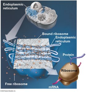
Ribosomes: Protein synthesis; can be free in the cytosol or bound to the endoplasmic reticulum (ER).
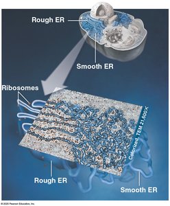
Endoplasmic Reticulum (ER): Network of membranes; rough ER (with ribosomes) synthesizes proteins, smooth ER synthesizes lipids.
Golgi Apparatus: Modifies, sorts, and packages proteins and lipids for secretion or delivery to other organelles.
Lysosomes: Contain digestive enzymes to break down macromolecules and damaged organelles.
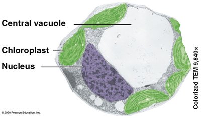
Vacuoles: Large vesicles for storage and transport; central vacuole in plants maintains turgor pressure.
Mitochondria: Sites of cellular respiration; generate ATP from organic molecules.
Chloroplasts (plants and algae): Sites of photosynthesis; convert solar energy to chemical energy.
Cytoskeleton: Network of microtubules, microfilaments, and intermediate filaments for structural support and transport.
Plasma Membrane: Controls entry and exit of substances.
Cell Wall (plants, fungi, some protists): Rigid structure outside the plasma membrane for support and protection.
Plasmodesmata (plants): Channels between adjacent plant cells for communication.
The Nucleus and Ribosomes
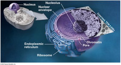
Nucleus
The nucleus is the genetic control center of eukaryotic cells.
Nuclear Envelope: Double membrane with pores for molecular exchange.
Nucleolus: Site of ribosomal RNA (rRNA) synthesis and ribosome assembly.
Chromatin: DNA-protein complex for efficient DNA packaging.
Ribosomes
Ribosomes are molecular machines that synthesize proteins using instructions from messenger RNA (mRNA).
Free Ribosomes: Float in the cytosol; synthesize proteins for use within the cell.
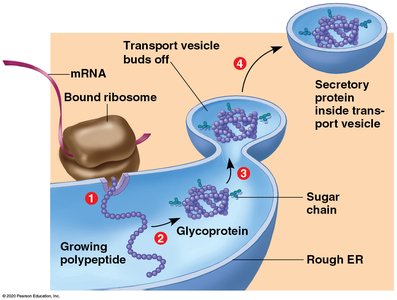
Bound Ribosomes: Attached to the rough ER; synthesize proteins for secretion or for use in membranes.
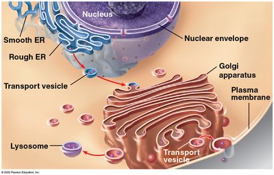
The Endomembrane System
Components and Functions
The endomembrane system is a group of interconnected organelles that work together to modify, package, and transport lipids and proteins.
Nuclear Envelope
Endoplasmic Reticulum (ER): Rough ER (protein synthesis), Smooth ER (lipid synthesis)
Golgi Apparatus: Modifies and sorts products from the ER
Lysosomes: Digestive compartments
Vesicles: Transport materials between organelles
Plasma Membrane: Exports products out of the cell
Endoplasmic Reticulum (ER)
Rough ER: Studded with ribosomes; synthesizes proteins for secretion or membrane insertion.
Smooth ER: Lacks ribosomes; synthesizes lipids, metabolizes carbohydrates, detoxifies drugs.
Protein Production in the ER
Proteins synthesized by ribosomes on the rough ER are often modified (e.g., glycosylation) and packaged into vesicles for transport to the Golgi apparatus.
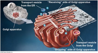
Golgi Apparatus
The Golgi apparatus consists of flattened membranous sacs (cisternae) and functions as the cell's shipping and receiving center.
Receives vesicles from the ER
Modifies proteins and lipids (e.g., adds carbohydrate groups)
Sorts and packages materials into vesicles for delivery

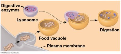
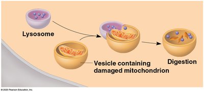
Lysosomes
Lysosomes are membrane-bound sacs containing hydrolytic enzymes for intracellular digestion.
Break down macromolecules, old organelles, and foreign substances
Play a key role in autophagy and phagocytosis
Vacuoles
Central Vacuole: Large organelle in plant cells for storage and maintaining turgor pressure.
Food Vacuole: Formed by phagocytosis in some protists and animal cells.
Mitochondria and Chloroplasts
Mitochondria
Mitochondria are the powerhouses of the cell, generating ATP through cellular respiration.
Outer Membrane: Smooth and protective
Inner Membrane: Folded into cristae to increase surface area
Matrix: Contains enzymes, mitochondrial DNA, and ribosomes
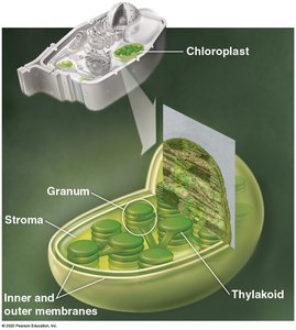
Chloroplasts
Chloroplasts are found in plants and algae and are the sites of photosynthesis.
Thylakoids: Flattened sacs where light reactions occur
Granum: Stack of thylakoids
Stroma: Fluid surrounding thylakoids; contains DNA, ribosomes, and enzymes
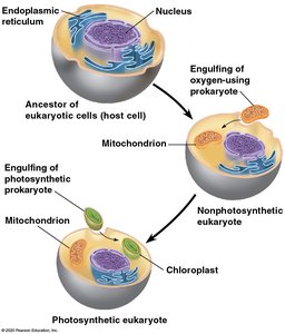
Endosymbiotic Theory
This theory proposes that mitochondria and chloroplasts originated as free-living prokaryotes that were engulfed by ancestral eukaryotic cells, forming a symbiotic relationship.
Both organelles have their own DNA and ribosomes
They replicate independently of the cell
Cytoskeleton and Cell Movement
Cytoskeleton
The cytoskeleton is a dynamic network of protein filaments that provides structural support, facilitates cell movement, and organizes organelles.
Microtubules: Hollow tubes made of tubulin; involved in cell shape, organelle movement, and chromosome separation
Intermediate Filaments: Rope-like fibers for mechanical strength
Microfilaments: Thin filaments of actin; involved in cell movement and shape
Cilia and Flagella
Cilia and flagella are motile appendages powered by the cytoskeleton, enabling movement or the transport of substances across cell surfaces.
Cilia: Short, numerous, move fluid or cells over a surface
Flagella: Longer, usually one or a few per cell, propel the cell
Extracellular Structures and Cell Junctions
Extracellular Matrix (ECM)
The ECM is a network of glycoproteins, polysaccharides, and proteoglycans outside animal cells, providing structural support and mediating cell signaling.
Collagen: Main structural protein in connective tissues
Integrins: Transmembrane proteins connecting ECM to the cytoskeleton
Cell Junctions
Cell junctions are specialized structures that connect adjacent cells and facilitate communication and adhesion.
Tight Junctions: Seal neighboring cells to prevent leakage of extracellular fluid
Desmosomes: Anchor cells together using intermediate filaments
Gap Junctions: Provide cytoplasmic channels for communication between animal cells
Plasmodesmata (plants): Channels through cell walls for transport and communication
Plant Cell Walls
Plant cell walls are rigid structures outside the plasma membrane, composed mainly of cellulose, providing support and protection.
Cellulose: A polysaccharide that forms the main component of plant cell walls
Plasmodesmata: Allow exchange of materials between plant cells
Summary Table: Prokaryotic vs. Eukaryotic Cells
Feature | Prokaryotic Cells | Eukaryotic Cells |
|---|---|---|
Nucleus | No (nucleoid region) | Yes (membrane-bound) |
Organelles | No membrane-bound organelles | Membrane-bound organelles present |
Size | Smaller (1-10 μm) | Larger (10-100 μm) |
Examples | Bacteria, Archaea | Animals, Plants, Fungi, Protists |
