Textbook Question
Receptors in the saccule and utricle provide sensations of
(a) Angular acceleration
(b) Hearing
(c) Vibration
(d) Gravity and linear acceleration and deceleration
1
views
 Verified step by step guidance
 3:44
3:44 4:14
4:14 6:14
6:14Receptors in the saccule and utricle provide sensations of
(a) Angular acceleration
(b) Hearing
(c) Vibration
(d) Gravity and linear acceleration and deceleration
The basic receptors in the internal ear are the
(a) Utricles
(b) Saccules
(c) Hair cells
(d) Supporting cells
(e) Ampullae
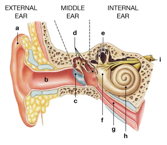
The malleus, incus, and stapes are the tiny bones located in the
(a) External ear
(b) Middle ear
(c) Internal ear
(d) Membranous labyrinth
Auditory information about the frequency and intensity of stimulation is relayed to the CNS over the cochlear nerve, a division of cranial nerve
(a) IV
(b) VI
(c) VIII
(d) X
The spiral organ is located in the_______of the internal ear.
(a) utricle
(b) bony labyrinth
(c) vestibule
(d) cochlea
Sound waves are converted into mechanical movements by the
(a) Auditory ossicles
(b) Cochlea
(c) Oval window
(d) Round window
(e) Tympanic membrane