Textbook Question
How does the heart's ability to contract differ from that of other muscles of the body?
1
views

 Verified step by step guidance
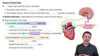
 6:13
6:13 02:31
02:31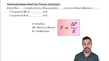 6:40
6:40How does the heart's ability to contract differ from that of other muscles of the body?
More than one choice may apply.
Which heart chamber pumps blood with the greatest amount of force?
a. Right atrium
b. Right ventricle
c. Left atrium
d. Left ventricle
Name three different factors that increase heart rate.
More than one choice may apply.
Using the figure, determine which vessels have the most resistance.
a. Arteries
b. Veins
c. Capillaries
d. Arterioles
e. Venules
More than one choice may apply.
How many cusps does the right atrioventricular valve have?
a. Two
b. Three
c. Four
d. Six
Trace one drop of blood from the time it enters the right atrium of the heart until it enters the left atrium. What is this circuit called?