 Back
BackBlood Vessels: Structure, Types, and Circulation
Study Guide - Smart Notes
 Tailored notes based on your materials, expanded with key definitions, examples, and context.
Blood Vessels
Overview of Blood Vessels
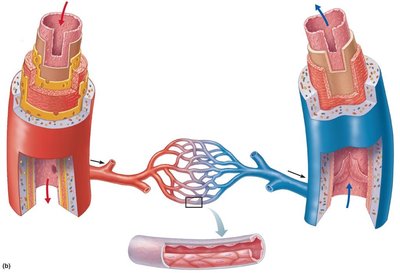
Blood vessels are essential components of the circulatory system, responsible for transporting blood throughout the body. There are three major types of blood vessels: arteries, capillaries, and veins. Each type has a unique structure and function, contributing to the efficient distribution and return of blood.
Arteries: Carry blood away from the heart.
Capillaries: Facilitate exchange of gases, nutrients, and wastes between blood and tissues.
Veins: Return blood to the heart.
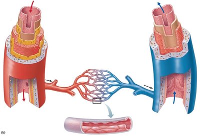
Structure of Blood Vessel Walls
General Organization
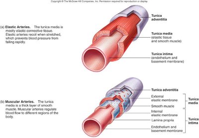
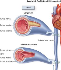
Most blood vessels (except capillaries) are composed of three layers, known as tunics:
Tunica intima: The innermost layer, consisting of endothelium (simple squamous epithelium) and, in larger vessels, a subendothelial layer.
Tunica media: The middle layer, made up of smooth muscle and elastic fibers. Responsible for vasoconstriction and vasodilation.
Tunica externa (adventitia): The outermost layer, composed of connective tissue that provides structural support and protection.
Lumen: The central blood-filled space within the vessel.
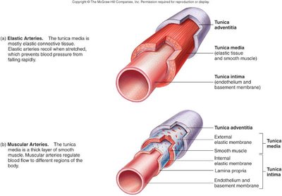
Comparison of Arterial Wall Structure
Arteries are classified based on the composition and thickness of their tunics:
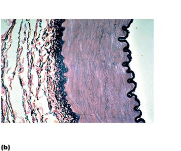
Elastic arteries: Largest arteries with a high elastin content in the tunica media, allowing them to withstand and dampen the pressure surges from the heart.
Muscular arteries: Medium-sized arteries with a thick tunica media composed mainly of smooth muscle, allowing for regulation of blood flow to different body regions.
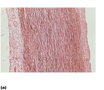
Types of Arteries
Elastic Arteries
Elastic arteries, such as the aorta and its major branches, have diameters ranging from 1 cm to 2.5 cm. Their high elastin content allows them to act as pressure reservoirs, maintaining blood flow during ventricular relaxation.
Muscular (Distributing) Arteries
Muscular arteries distribute blood to specific organs and regions. They have a thick tunica media and prominent internal and external elastic membranes.
Arterioles
Arterioles are the smallest arteries, with diameters from 10 µm to 0.3 mm. They regulate blood flow into capillary beds through vasoconstriction and vasodilation, influenced by local tissue factors and the sympathetic nervous system.
Capillaries
Structure and Function
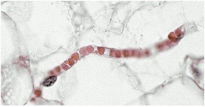
Capillaries are the smallest blood vessels (8–10 µm in diameter), allowing red blood cells to pass through in single file. They are the primary sites for exchange of gases, nutrients, and wastes between blood and tissues.
Lungs: Oxygen enters blood, carbon dioxide leaves.
Small intestines: Absorb digested nutrients.
Endocrine glands: Pick up hormones.
Kidneys: Remove nitrogenous wastes.
Types of Capillaries
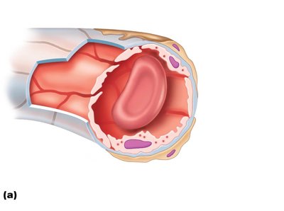
Continuous capillaries: Most common; have tight junctions and intercellular clefts for limited permeability.
Fenestrated capillaries: Contain pores (fenestrations) for increased permeability; found in kidneys, small intestine, and endocrine glands.
Sinusoids: Wide, leaky capillaries with large intercellular clefts; found in liver, bone marrow, and spleen.
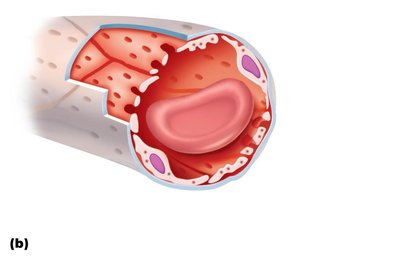
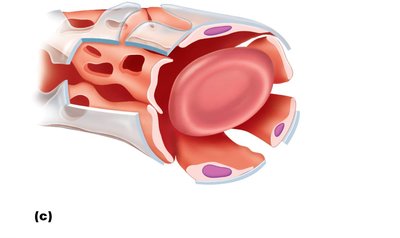
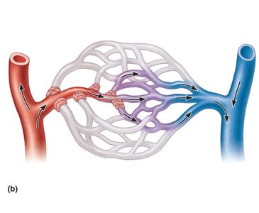
Capillary Beds and Blood Flow Regulation
Capillary beds are networks of capillaries within tissues. Blood flow is regulated by precapillary sphincters, which open or close to direct blood through true capillaries or shunt it through a vascular channel.
Veins and Venous Return
Structure and Function
Veins return blood to the heart and have several structural differences from arteries:
Larger lumens and thinner walls.
Thicker tunica externa and less elastin.
Contain valves, especially in limbs, to prevent backflow.
Blood pressure is much lower than in arteries.
Major Circulatory Pathways
Pulmonary Circulation
The pulmonary circuit carries blood between the heart and lungs for gas exchange. The pulmonary trunk leaves the right ventricle, dividing into right and left pulmonary arteries, while pulmonary veins return oxygenated blood to the left atrium.
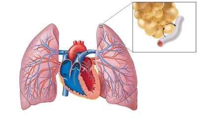
Systemic Circulation
The systemic circuit delivers oxygenated blood from the heart to the body and returns deoxygenated blood to the heart. The aorta is the main artery, branching into arteries that supply the head, trunk, and limbs.
Disorders of Blood Vessels
Common Vascular Disorders
Aneurysm: Localized dilation of a blood vessel wall, often in the aorta, which can rupture if untreated.
Deep vein thrombosis: Formation of blood clots in deep veins, usually in the lower limbs.
Venous disease: Includes varicose veins and chronic venous insufficiency.
Microangiopathy of diabetes: Damage to small blood vessels due to chronic high blood sugar.
Arteriovenous malformation: Abnormal connection between arteries and veins, bypassing capillaries.
Summary Table: Comparison of Arteries, Capillaries, and Veins
Feature | Arteries | Capillaries | Veins |
|---|---|---|---|
Direction of Blood Flow | Away from heart | Between arteries and veins | Toward heart |
Wall Thickness | Thick | Very thin (one cell layer) | Thin |
Valves | Absent | Absent | Present (especially in limbs) |
Pressure | High | Low | Lowest |
