 Back
BackBlood Vessels: Structure, Function, and Circulation
Study Guide - Smart Notes
 Tailored notes based on your materials, expanded with key definitions, examples, and context.
Blood Vessels
Types of Blood Vessels
Blood vessels form a closed delivery system that transports blood throughout the body. There are three major types of blood vessels:
Arteries: Carry blood away from the heart.
Capillaries: The smallest blood vessels, where exchange of substances occurs.
Veins: Carry blood toward the heart.
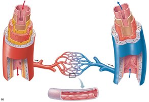
Arteries branch into smaller arterioles, which lead to capillaries. Capillaries then merge into venules, which join to form veins.
Structure of Blood Vessel Walls
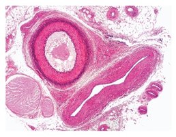
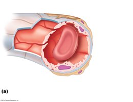
Blood vessel walls are composed of three layers, or tunics:
Tunica intima: The innermost layer, consisting of endothelium (simple squamous epithelium) and, in larger vessels, a subendothelial layer.
Tunica media: The middle layer, made up of smooth muscle and elastic fibers. Responsible for vasoconstriction (contraction) and vasodilation (relaxation).
Tunica externa: The outermost layer, composed of connective tissue (mainly collagen fibers).
The lumen is the central blood-filled space of a vessel.
Types of Arteries
Elastic Arteries
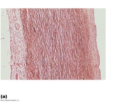
Elastic arteries are the largest arteries (diameter 1–2.5 cm), including the aorta and its major branches. They are also called conducting arteries due to their high elastin content, which helps dampen the surge of blood pressure from the heart.
Muscular (Distributing) Arteries
Muscular arteries lie distal to elastic arteries and have diameters ranging from 0.3 cm to 1 mm. They have a thick tunica media and unique internal and external elastic membranes. Most named arteries are muscular arteries.
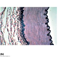
Arterioles
Arterioles are the smallest arteries (diameter 10 μm to 0.3 mm). Larger arterioles possess all three tunics. Their diameter is regulated by local tissue factors and the sympathetic nervous system.
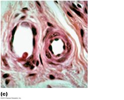
Capillaries
Structure and Function
Capillaries are the smallest blood vessels (diameter 8–10 μm), allowing red blood cells to pass through single file. They are the primary site for exchange of gases, nutrients, and wastes between blood and tissues. Capillaries have site-specific functions:
Lungs: Gas exchange (O2 in, CO2 out)
Small intestine: Absorption of nutrients
Endocrine glands: Pick up hormones
Kidneys: Removal of nitrogenous wastes
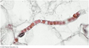
Types of Capillaries
Continuous capillaries: Most common, with tight junctions and intercellular clefts allowing limited passage of small molecules.
Fenestrated capillaries: Have pores (fenestrations) that increase permeability, found in areas of active absorption or filtration (e.g., kidneys, small intestine).
Sinusoids: Wide, leaky capillaries with large intercellular clefts, found in bone marrow, liver, and spleen.
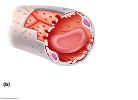
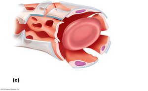
Capillary Permeability
Substances move into and out of capillaries by four routes:
Direct diffusion through endothelial cell membranes
Intercellular clefts
Fenestrations (pores)
Pinocytotic vesicles
Capillary Beds
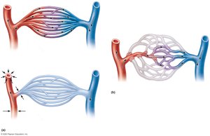
Capillary beds are networks of capillaries running through tissues. Blood flow is regulated by precapillary sphincters, which open and close to direct blood flow according to tissue needs.
Veins and Venous Return
Venous Vessels
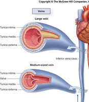
Veins conduct blood from capillaries back toward the heart. Blood pressure in veins is much lower than in arteries. Venules are the smallest veins, joining to form larger veins. Veins have larger lumens, thicker tunica externa, less elastin, and thinner walls compared to arteries.
Venous Valves and Skeletal Muscle Pump
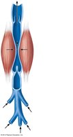
To counteract low venous pressure and prevent backflow, veins (especially in the limbs) contain valves. The skeletal muscle pump assists venous return by compressing veins during muscle contraction.
Pulmonary and Systemic Circulation
Pulmonary Circulation
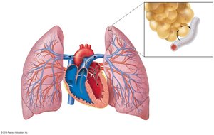
The pulmonary circuit carries blood between the heart and lungs. The pulmonary trunk leaves the right ventricle and divides into right and left pulmonary arteries. Pulmonary veins return oxygenated blood to the left atrium. Pulmonary vessels have thinner walls and lower pressure than systemic vessels.
Systemic Circulation
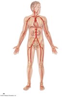
Systemic arteries carry oxygenated blood from the heart to the body. The aorta is the largest artery, branching into major arteries that supply the head, trunk, and limbs.
Major Arteries and Veins
The Aorta and Its Branches
The aorta has three main parts: ascending aorta, aortic arch, and descending aorta. Major branches supply the head, neck, upper limbs, and lower body.
Arteries of the Head, Neck, and Brain
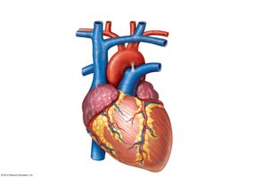
The common carotid arteries branch into internal and external carotid arteries, supplying the brain and face. The vertebral arteries supply the posterior brain and join to form the basilar artery, contributing to the cerebral arterial circle (circle of Willis).
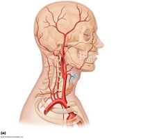
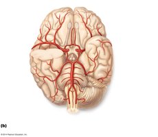
Arteries of the Upper Limb
The axillary artery becomes the brachial artery, which divides into the radial and ulnar arteries supplying the forearm and hand.
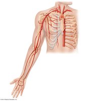
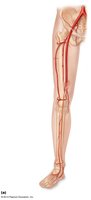
Arteries of the Abdomen and Lower Limbs
The abdominal aorta gives rise to several branches supplying abdominal organs and lower limbs, including the celiac trunk, mesenteric arteries, and common iliac arteries.
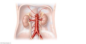
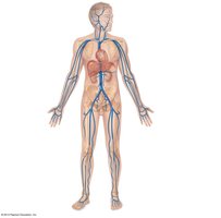
Major Veins and Venous Return
The superior and inferior vena cava return blood to the right atrium from the upper and lower body, respectively. Major veins include the jugular, subclavian, brachiocephalic, and iliac veins.
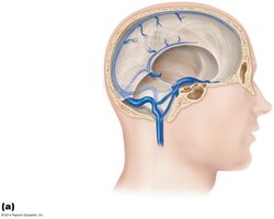
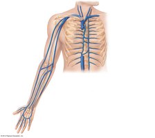
Veins of the Head, Neck, and Upper Limb
Dural venous sinuses drain the brain, while internal and external jugular veins drain the head and neck. Superficial veins of the upper limb include the cephalic, basilic, and median cubital veins.
Veins of the Abdomen
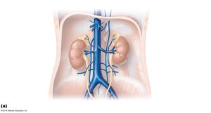
Major abdominal veins include the renal, hepatic, and gonadal veins, which drain into the inferior vena cava.
The Hepatic Portal System
Function and Structure
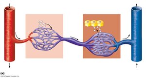
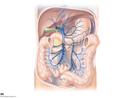
The hepatic portal system is a specialized vascular circuit that transports blood from the digestive organs to the liver for processing. It consists of two capillary beds: one in the digestive tract and one in the liver (liver sinusoids).
Disorders of Blood Vessels
Common Disorders
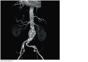
Aneurysm: A localized dilation of a blood vessel wall, often in the aorta, which can rupture and cause life-threatening bleeding.
Deep vein thrombosis: Formation of a blood clot in a deep vein, usually in the lower limb.
Venous disease: Includes varicose veins and chronic venous insufficiency.
Microangiopathy of diabetes: Damage to small blood vessels due to chronic high blood sugar.
Arteriovenous malformation: Abnormal connection between arteries and veins, bypassing capillaries.
Blood Vessels in Adulthood
Atherosclerosis, the buildup of fatty plaques in arteries, begins in youth but its consequences become evident in middle to old age. Males are more affected between ages 45 and 65, but females experience heart disease and atherosclerosis later in life.
