What is the formula for calculating heart rate using the QRS complexes?
Heart rate can be calculated by counting the number of QRS complexes in a given time frame (usually 60 seconds) and multiplying by a factor to convert to beats per minute.
How are cardiac muscle cells connected?
Cardiac muscle cells are connected by gap junctions
Starting at the right atrium, what is the sequence of blood flow through the heart?
Blood flows from the right atrium to the right ventricle, then to the pulmonary arteries, lungs, pulmonary veins, left atrium, left ventricle, and finally to the aorta.
What occurs during the QRS complex?
The QRS complex represents ventricular depolarization, which leads to ventricular contraction.
Which wave on an ECG represents atrial depolarization?
P wave
Which structure is not part of the intrinsic conduction system of the heart?
Coronary arteries
Which medication reduces the workload of the heart by treating abnormal heart rhythms?
Beta-blockers reduce the workload of the heart by treating abnormal heart rhythms.
Which vessels receive blood during ventricular systole?
Pulmonary arteries
What is the priority intervention for a patient in ventricular fibrillation?
The priority intervention for ventricular fibrillation is defibrillation.
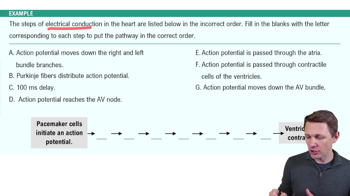
During a heartbeat, what is the role of the atrioventricular (AV) node?
The AV node delays the electrical impulse to allow the ventricles to fill with blood before they contract.
Why are gap junctions a vital part of the intercellular connection of cardiac muscles?
Gap junctions allow for the rapid spread of action potentials between cardiac muscle cells, enabling synchronized contraction.
How is cardiac output calculated?
Cardiac output is calculated as the product of heart rate and stroke volume.
Which wave on an ECG represents ventricular depolarization?
QRS complex
Which mineral is especially important for maintaining proper heart rhythm?
Potassium
What is the correct sequential path of an action potential in the heart?
SA node, AV node, Bundle of His, Purkinje fibers
How do the ventricles fill with blood during the cardiac cycle?
The ventricles fill with blood during diastole, primarily due to passive flow from the atria.
What is the correct formula for calculating cardiac output?
Heart rate x Stroke volume
Which structures coordinate contraction in cardiac muscle cells?
Gap junctions and conducting fibers coordinate contraction in cardiac muscle cells.
Which part of the cardiac conduction system receives electrical impulses from the bundle branches?
The Purkinje fibers receive electrical impulses from the bundle branches.
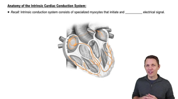
What is true concerning the heart conduction system's function?
It initiates and conducts action potentials autonomously
Which part of the waveform represents atrioventricular conduction?
The PR interval represents atrioventricular conduction.
Where is the SA node located?
The SA node is located in the superior right atrial wall, near the entrance of the superior vena cava.
What controls heartbeat?
The heartbeat is controlled by the intrinsic cardiac conduction system, primarily the SA node.
Which structure is not part of the cardiac conduction system?
Coronary arteries
What indicates ventricular depolarization on an ECG?
QRS complex
A client is administered atropine to increase the heart rate. What is the action of atropine?
Atropine blocks the parasympathetic influence on the heart, increasing heart rate.
A nurse is preparing a client for cardiac catheterization. What is the nurse’s priority assessment?
The nurse's priority assessment is to check for allergies, especially to iodine or shellfish, due to the use of contrast dye.
What is another name for the pacemaker potential?
Prepotential
What initiates the release of calcium from the sarcoplasmic reticulum in cardiac muscle cells?
The influx of calcium through voltage-gated calcium channels during depolarization initiates the release of calcium from the sarcoplasmic reticulum.
What are the key considerations for cardiac catheterization? (Select all that apply.) A) Check for allergies B) Monitor vital signs C) Ensure fasting D) Administer sedatives
A) Check for allergies, B) Monitor vital signs, C) Ensure fasting
Which description of cardiac pacemaker cells is incorrect?
They require neural input to function
Which of these statements about the heart is true? A) The heart requires external signals to beat B) The heart's rhythm is set by the AV node C) The heart can beat independently of the nervous system D) The heart's contraction is initiated by skeletal muscle
C) The heart can beat independently of the nervous system
Which part of the waveform represents ventricular depolarization? A) P wave B) QRS complex C) T wave D) U wave
B) QRS complex
In which situation should you avoid administering a synchronized shock?
Ventricular fibrillation
Where are the cardiac conduction fibers located?
Cardiac conduction fibers are located throughout the heart, including the SA node, AV node, bundle of His, bundle branches, and Purkinje fibers.
How is backflow of blood from the right ventricle into the right atrium prevented?
The tricuspid valve prevents backflow of blood from the right ventricle into the right atrium.
How should a nurse assess a newborn's heart rate?
The nurse should use a stethoscope to auscultate the apical pulse for a full minute to assess the newborn's heart rate.
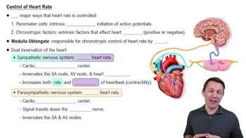
Where is the cardiac center located?
The cardiac center is located in the medulla oblongata of the brainstem.
Which is most responsible for the synchronized contraction of cardiac muscle tissue?
Gap junctions are most responsible for the synchronized contraction of cardiac muscle tissue.
 Back
Back
 6:13
6:13