Ch. 19 - Amines
Wade - Organic Chemistry 9th Edition
Wade9th EditionOrganic ChemistryISBN: 9780135213728Not the one you use?Change textbook
Chapter 19, Problem 41

The mass spectrum of tert-butylamine follows shows an intense base peak at m/z 58, and very little else. Use a diagram to show the cleavage that accounts for the base peak. Suggest why no molecular ion is visible in this spectrum.

Verified step by step guidance
1
Analyze the mass spectrum provided. The base peak at m/z 58 indicates the most stable and abundant fragment ion formed during the fragmentation of tert-butylamine.
Understand the structure of tert-butylamine: (CH3)3CNH2. The molecule consists of a tert-butyl group ((CH3)3C-) attached to an amine (-NH2). Fragmentation typically occurs at the weakest bond or the most favorable cleavage site.
Propose the cleavage mechanism: The bond between the tert-butyl group and the amine group is likely to break, forming a stable carbocation (C4H10+, m/z 58). This carbocation is stabilized by hyperconjugation and inductive effects from the three methyl groups.
Explain the absence of a molecular ion peak: The molecular ion (m/z 73 for tert-butylamine) is likely absent due to its instability. Tert-butylamine undergoes rapid fragmentation upon ionization, favoring the formation of the stable m/z 58 fragment instead of retaining the molecular ion.
Draw a diagram to illustrate the cleavage: Show the tert-butylamine structure, the bond cleavage between the tert-butyl group and the amine group, and the formation of the stable carbocation (C4H10+, m/z 58). This visual representation helps clarify the fragmentation process.

Verified video answer for a similar problem:

This video solution was recommended by our tutors as helpful for the problem above.
Video duration:
6m

Key Concepts

Here are the essential concepts you must grasp in order to answer the question correctly.

Mass Spectrometry

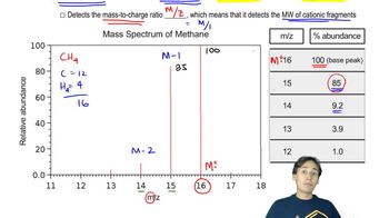
Mass spectrometry is an analytical technique used to measure the mass-to-charge ratio of ions. It involves ionizing chemical species and sorting the resulting ions based on their mass. The output is a mass spectrum, which displays the relative abundance of detected ions at various mass-to-charge (m/z) ratios, providing insights into the molecular structure and composition of the sample.
Recommended video:
01:25
How to Read a Mass Spectrum

Base Peak

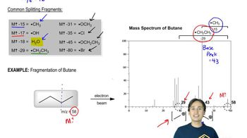
The base peak in a mass spectrum is the peak with the highest intensity, representing the most abundant ion detected. In the case of tert-butylamine, the base peak at m/z 58 indicates a stable fragment ion resulting from the cleavage of the molecule. Understanding the base peak helps in identifying the most significant fragmentation pathways and the structure of the compound.
Recommended video:
01:27
Clarification on the Base Peak

Molecular Ion

The molecular ion is the ion that corresponds to the intact molecule of the analyte, typically represented as M+. Its absence in a mass spectrum can indicate that the molecule has fragmented before detection, often due to instability or the presence of weak bonds. In tert-butylamine's spectrum, the lack of a molecular ion suggests that fragmentation occurs readily, leading to the formation of smaller, more stable ions.
Recommended video:
Guided course
3:39
Metal Ion Catalysis Concept 1