What is the purpose of special staining techniques in microbiology?
Special staining techniques are used to visualize specific cell structures, either inside or outside cells, that are not easily seen with standard stains.
What structure does the capsule stain highlight in bacteria?
The capsule stain highlights the capsule surrounding bacteria, making it visible as a white border.
How do endospores appear when stained in Bacillus cereus?
Endospores appear as bluish-green dots when stained in Bacillus cereus.
Why are flagella stains necessary for observing flagella?
Flagella stains are necessary because flagella are not visible without this specific staining technique.
What is the main advantage of using special stains like capsule, endospore, and flagella stains?
They allow scientists to easily visualize specific structures of interest that are otherwise difficult to see.
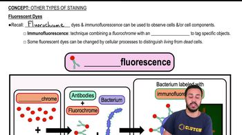
What is immunofluorescence used for in microbiology?
Immunofluorescence is used to tag specific molecules or antigens on cells by combining fluorescent dyes with antibodies.
How does immunofluorescence indicate the presence of specific antigens?
The fluorescence from the bound antibodies indicates the presence of specific antigens on the surface of bacteria.
What are fluorochrome dyes, and what is their role in cell observation?
Fluorochrome dyes are fluorescent molecules used to observe cells and their components by emitting fluorescence when bound to specific targets.
How can some fluorescent dyes differentiate between living and dead cells?
Some fluorescent dyes are changed by cellular processes, allowing them to distinguish living cells (with active processes) from dead cells.
What is the function of antibodies in immunofluorescence staining?
Antibodies bind to specific antigens on cells, enabling targeted staining when combined with fluorescent dyes.
What color are the fluorochrome molecules depicted in the immunofluorescence diagram?
The fluorochrome molecules are depicted as red circles in the diagram.
What shape are antibodies typically represented as in immunofluorescence diagrams?
Antibodies are typically represented as Y-shaped proteins in immunofluorescence diagrams.
What happens to the fluorochrome when the antibody binds to the antigen?
The fluorochrome fluoresces, and this fluorescence can be detected, indicating antigen presence.
Why are flagella not visible in the capsule stain image of Bacillus cereus?
Flagella are not visible because the flagella stain is not used in the capsule stain image.
What is an antigen in the context of immunofluorescence?
An antigen is a molecule on the cell surface that antibodies specifically bind to during immunofluorescence.
 Back
Back
 02:59
02:59