Textbook Question
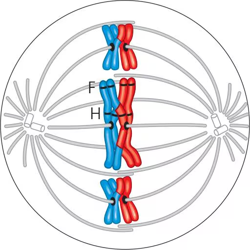
The following diagram shows a cell in meiosis. Identify the stage of meiosis shown.
 Verified step by step guidance
 04:49
04:49 08:09
08:09 05:08
05:08The following diagram shows a cell in meiosis. Identify the stage of meiosis shown.
If the DNA content of a diploid cell in the G1 phase of the cell cycle is x, then the DNA content of the same cell at metaphase of meiosis I will be
a. 0.25x
b. 0.5x
c. x
d. 2x
Meiosis II is similar to mitosis in that
a. Sister chromatids separate during anaphase
b. DNA replicates before the division
c. The daughter cells are diploid
d. Homologous chromosomes synapse
If we continue to follow the cell lineage from question 4, then the DNA content of a single cell at metaphase of meiosis II will be
a. 0.25x
b. 0.5x
c. x.
d. 2x
Assume that genes A and B are on the same chromosome and are 50 map units apart. An animal heterozygous at both loci is crossed with one that is homozygous recessive at both loci. What percentage of the offspring will show recombinant phenotypes resulting from crossovers? Without knowing these genes are on the same chromosome, how would you interpret the results of this cross?
The diagram shows a cell in meiosis. Describe the makeup of a haploid set and a diploid set.