Textbook Question
The point of the elbow is actually the _____ of the ulna.
(a) Styloid process
(b) Olecranon
(c) Coronoid process
(d) Trochlear notch
2
views

 -+
-+ Verified step by step guidance
 02:40
02:40 3:00
3:00 06:04
06:04The point of the elbow is actually the _____ of the ulna.
(a) Styloid process
(b) Olecranon
(c) Coronoid process
(d) Trochlear notch
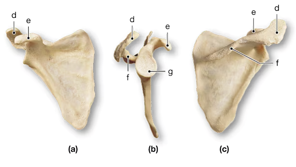
Which of the following is primarily responsible for stabilizing, positioning, and bracing the pectoral girdle?
(a) Tendons
(b) Ligaments
(c) The joint shape
(d) Muscles
(e) The shape of the bones within the joint
In anatomical position, the ulna lies:
(a) Medial to the radius
(b) Lateral to the radius
(c) Inferior to the radius
(d) Superior to the radius