Ch. 4 The Tissue Level of Organization
Martini - Fundamentals of Anatomy & Physiology 11th Edition
Martini, Nath, Bartholomew11th EditionFundamentals of Anatomy & PhysiologyISBN: 9780136874089Not the one you use?Change textbook
Chapter 4, Problem 1e

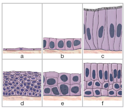
Identify the six types of epithelial tissue shown in the drawing below.
e. ___
Illustration of six types of epithelial tissue cells labeled a to f, showing various shapes and arrangements.

Verified step by step guidance
1
Step 1: Identify the shape and layering of the cells in each image. Epithelial tissues are classified based on the number of cell layers (simple = one layer, stratified = multiple layers) and the shape of the cells (squamous = flat, cuboidal = cube-shaped, columnar = tall and column-like).
Step 2: For the first image (top left), observe that the cells are flat and arranged in a single layer. This corresponds to 'Simple Squamous Epithelium'.
Step 3: For the second image (top middle), the cells are cube-shaped and arranged in a single layer. This corresponds to 'Simple Cuboidal Epithelium'.
Step 4: For the third image (top right), the cells are tall and column-like, arranged in a single layer. This corresponds to 'Simple Columnar Epithelium'.
Step 5: For the fourth image (bottom left), multiple layers of flat cells are present. This corresponds to 'Stratified Squamous Epithelium'. For the fifth image (bottom middle), multiple layers of cube-shaped cells are present, corresponding to 'Stratified Cuboidal Epithelium'. For the sixth image (bottom right), multiple layers of columnar cells are present, corresponding to 'Stratified Columnar Epithelium'.

Verified video answer for a similar problem:

This video solution was recommended by our tutors as helpful for the problem above.
Video duration:
3m
Was this helpful?

Key Concepts

Here are the essential concepts you must grasp in order to answer the question correctly.

Types of Epithelial Tissue

Epithelial tissues are classified based on cell shape and layering. The six main types include simple squamous, simple cuboidal, simple columnar, stratified squamous, stratified cuboidal, and pseudostratified columnar. Each type has distinct structural features and functions related to protection, absorption, or secretion.
Recommended video:
Guided course
07:25
Identifying Types of Epithelial Tissue Example 1

Cell Shape and Arrangement

Epithelial cells are categorized by their shape—squamous (flat), cuboidal (cube-shaped), or columnar (tall and column-like). The arrangement can be simple (single layer), stratified (multiple layers), or pseudostratified (appears layered but all cells contact the basement membrane). These characteristics determine the tissue's role and location.
Recommended video:
Guided course
8:11
Fascicle Arrangements

Function and Location of Epithelial Types

Each epithelial type serves specific functions: simple squamous facilitates diffusion, simple cuboidal aids secretion and absorption, simple columnar specializes in absorption and secretion, stratified squamous protects against abrasion, stratified cuboidal provides protection in ducts, and pseudostratified columnar often has cilia for moving mucus. Understanding these helps identify tissue types in the body.
Recommended video:
Guided course
07:25
Identifying Types of Epithelial Tissue Example 1