Ch. 20 The Heart
Martini - Fundamentals of Anatomy & Physiology 11th Edition
Martini, Nath, Bartholomew11th EditionFundamentals of Anatomy & PhysiologyISBN: 9780136874089Not the one you use?Change textbook
Chapter 20, Problem 12

What role do the chordae tendineae and papillary muscles play in the normal function of the AV valves?

Verified step by step guidance
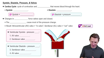
1
Understand that the atrioventricular (AV) valves, which include the tricuspid and mitral valves, prevent backflow of blood from the ventricles to the atria during ventricular contraction.
Recognize that the chordae tendineae are strong, fibrous strings attached to the edges of the AV valve leaflets and anchored to the papillary muscles located on the inner walls of the ventricles.
Know that during ventricular contraction (systole), the papillary muscles contract, which pulls on the chordae tendineae, creating tension that holds the valve leaflets in a closed position.
This tension prevents the valve leaflets from prolapsing or inverting back into the atria, ensuring a one-way flow of blood and maintaining efficient heart function.
Summarize that the chordae tendineae and papillary muscles work together to stabilize the AV valves during ventricular contraction, preventing valve regurgitation and maintaining proper cardiac circulation.

Verified video answer for a similar problem:

This video solution was recommended by our tutors as helpful for the problem above.
Was this helpful?

Key Concepts

Here are the essential concepts you must grasp in order to answer the question correctly.

Atrioventricular (AV) Valves

The AV valves, including the mitral and tricuspid valves, regulate blood flow between the atria and ventricles. They open to allow blood to flow into the ventricles and close to prevent backflow during ventricular contraction, ensuring unidirectional blood flow.
Recommended video:
Guided course
7:13
Systole, Diastole, Pressure, & Valves

Chordae Tendineae

Chordae tendineae are strong, fibrous cords that connect the AV valve leaflets to the papillary muscles. They prevent the valve leaflets from inverting or prolapsing into the atria during ventricular contraction, maintaining valve integrity and preventing regurgitation.

Papillary Muscles

Papillary muscles are muscular projections from the ventricular walls that anchor the chordae tendineae. During ventricular contraction, they contract simultaneously to tighten the chordae tendineae, stabilizing the AV valves and preventing valve prolapse.
Recommended video:
9:42
Types of Capillaries