Skip to main content
Anatomy & Physiology
My Course
Learn
Exam Prep
AI Tutor
Study Guides
Textbook Solutions
Flashcards
Explore
Try the app
My Course
Learn
Exam Prep
AI Tutor
Study Guides
Textbook Solutions
Flashcards
Explore
Try the app
Back
Vascular Layer of the Eyeball definitions
You can tap to flip the card.
Uvea
You can tap to flip the card.
👆
Uvea
The middle layer of the eye, consisting of the iris, ciliary body, and choroid, responsible for regulating light and supplying blood.
Track progress
Control buttons has been changed to "navigation" mode.
1/15
Related flashcards
Related practice
Recommended videos
Vascular Layer of the Eyeball quiz
Vascular Layer of the Eyeball
10 Terms
Vascular Layer of the Eyeball quiz #1
Vascular Layer of the Eyeball
11 Terms
Vascular Layer of the Eyeball
15. The Special Senses
1 problem
Topic
Optic Components of the Eyeball
15. The Special Senses
1 problem
Topic
Bruce
15. The Special Senses
8 topics
13 problems
Chapter
Bruce
Guided course
8:07
Control of Light Entering the Eye
Guided course
6:55
Vascular Layer
1
views
Guided course
3:56
Vascular Layer of the Eyeball Example 1
2
views
Terms in this set (15)
Hide definitions
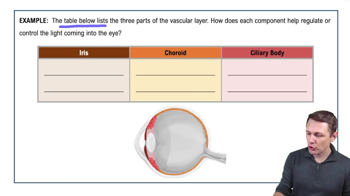
Uvea
The middle layer of the eye, consisting of the iris, ciliary body, and choroid, responsible for regulating light and supplying blood.
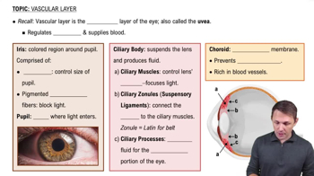
Iris
The colored part of the eye, controlling light entry by adjusting pupil size through muscles and pigmented elastic fibers.
Ciliary Body
A structure that suspends the lens, produces aqueous humor, and adjusts lens shape for focusing light.
Choroid
A pigmented membrane rich in blood vessels, located between the sclera and retina, preventing light reflection inside the eye.
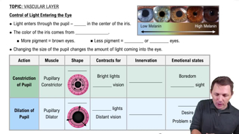
Pupil
A hole in the center of the iris that allows light to enter the eye, with its size controlled by the iris.
Melanin
A brown pigment in the iris determining eye color, with higher levels resulting in darker eyes.
Ciliary Muscles
Muscles that control the lens's shape, affecting light focus by making the lens rounder or flatter.
Ciliary Zonules
Ligaments connecting the ciliary muscles to the lens, aiding in lens shape adjustment for focusing.
Ciliary Processes
Structures that secrete aqueous humor, the fluid filling the front part of the eye.
Aqueous Humor
Fluid produced by the ciliary processes, filling the front part of the eye and maintaining intraocular pressure.
Pupillary Constrictor Muscle
A circular muscle in the iris that reduces pupil size in bright light or during close vision.
Pupillary Dilator Muscle
Radial muscles in the iris that increase pupil size in dim light or during distant vision.
Autonomic Nervous System
Regulates involuntary actions like pupil size, with parasympathetic causing constriction and sympathetic causing dilation.
Sympathetic Division
Part of the autonomic nervous system that dilates the pupil during fight or flight responses.
Parasympathetic Division
Part of the autonomic nervous system that constricts the pupil during rest and digest states.
 BackBack
 8:07
8:07